Thane: Shop No 10, 13 And 14, Upper Ground Floor, JVMS Corner Stone, Hari Niwas Circle, Naupada, Lbs Marg, Thane (West) - 400602     Nashik: Sharanpur Road, Behind Kulkarni Garden, Racca Colony, Jejurkar Wadi, Sharanpur, Nashik, Maharashtra 422002     Yeola: Yeola-Nashik Rd, Angan, Yeola, Maharashtra 423401

Email Id.

panaceahospital16@gmail.com

Emergency No.

0253-2946969 | 8958956996

Hysteroscopy

Hysteroscopy

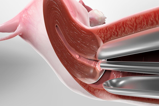
Hysteroscopy is a minimally invasive diagnostic and therapeutic procedure that allows doctors to view the inside of the uterus using a thin, lighted instrument called a hysteroscope. It helps in identifying and treating uterine problems such as fibroids, polyps, adhesions, or abnormal bleeding, thereby improving fertility and overall reproductive health.

When to Opt for Hysteroscopy?

  • For evaluation of recurrent miscarriages or infertility
  • In cases of abnormal uterine bleeding
  • To remove fibroids, polyps, or uterine adhesions
  • When congenital uterine abnormalities are suspected
  • Before IVF treatment to ensure a healthy uterine environment
  • If advised by a gynecologist after abnormal ultrasound findings

Precautions During the Procedure

  • Performed under proper anesthesia (local or general)
  • Sterile environment is maintained to prevent infections
  • Patients should avoid food and drink before the procedure if anesthesia is planned
  • Antibiotics may be prescribed in certain cases to reduce infection risk

Post-Treatment Care

  • Mild cramping or spotting is common for a few days
  • Avoid heavy exercise or lifting for 2–3 days
  • Abstain from sexual intercourse until advised by the doctor
  • Report any severe pain, fever, or heavy bleeding immediately
  • Follow-up visit is essential to review results and further treatment

Risks Involved

  • Mild cramping or uterine discomfort
  • Rare risk of infection or uterine perforation
  • Possible allergic reaction to anesthesia
  • Excessive bleeding in rare cases

Steps in Hysteroscopy

  • Preparation: Patient is given anesthesia or sedation as required.
  • Insertion: A hysteroscope is gently inserted into the uterus through the vagina and cervix.
  • Visualization: Saline or CO₂ gas is used to expand the uterus for clear viewing.
  • Diagnosis/Treatment: Any abnormalities like fibroids or polyps are identified and removed if necessary.
  • Completion: The hysteroscope is removed, and recovery begins.

This safe and minimally invasive procedure plays a crucial role in diagnosing and treating uterine conditions, improving fertility outcomes.

Advanced Technology

Modern hysteroscopes with high-definition imaging and miniaturized instruments allow for precise diagnosis and treatment with minimal discomfort and faster recovery.

Expert Consultation